Counter-stimulating Pain
(Workbook Pages 11,12, 13)
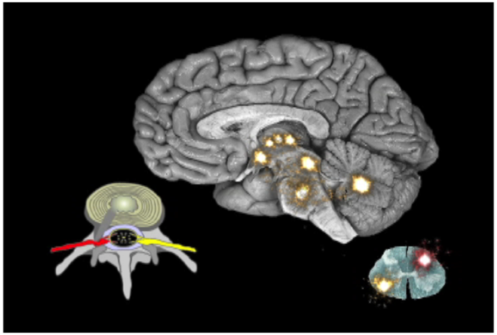
The graphics on page 10, 11, 12 and 13 are some of the most important ones in the Neuroplastic Transformation workbook. On page 10 we see the 2 regions in the spinal cord and 16 regions of the brain that make up the pain circuit. This depicts acute pain where 5% of the cells in each region are dedicated to pain processing. These nerve cells are reserved for pain and are shut off, until signaled by sensory pathways in the body to turn on due to danger in the form of injury, illness or inflammation. These signals are processed from the back part of the spinal cord, crossing over to the opposite side of the cord and sent up pathways to the brain stem and mid-brain. We only perceive the signal as pain after it reaches the thinking brain. Here in the 9 regions of thinking brain where the signal is received we experience it as pain. In acute pain some of the same regions and a few different ones send a signal back down to the spinal cord to intercept the incoming signal and shut off the pain so that it never reaches that upper part of the brain, which we use to perceive sensations. On page 11 we see the graphic that shows the same regions in the spinal cord and the brain being simulated, but this time the signal covers a greater area. This represents the recruitment of other nerve cells to expand the pain map and steal away cells from other regional processes. This is a basic principle of neuroplasticity. The brain assigns cells to specific functions, but if one function becomes more active, cells are taken from other functions to strengthen the more active one
Look at these two pictures on page 10 and 11, turning the pages back and forth. The picture on page 11 shows one of the key reasons why persistent pain is no longer a symptom, but has transformed to a disease. This is not because the pain is chronic, but is that expansion of brain real-estate to 15% to 25% of the cells in these regions that are processing the pain signals. Now look at the graphic on page 12. Here we see that even in wound-up and persistent signaling below the upper 9 regions of the thinking brain, if the signal does not advance into the thinking brain, there can be no pain. Turn page 11 and 12 back and forth and recognize that if the signal stops below the upper 9 regions of the brain, there can be no pain. Keep looking at the graphics on page 11 and 12 until you can memorize the difference and visualize them without the actual pictures. Remind yourself that if your brain looks like the graphic on page 12 you cannot have pain.